


Choosing the Right Doctor and Procedure For Your Spine
When it comes to back surgery, patients in the Los Angeles area have many choices. Today, lumbar discectomy and spinal fusion procedures can be performed through tiny incisions as out-patient procedures. With so many choices, how does a patient choose a procedure or surgeon?
It’s important to know the qualifications, training and experience of the Doctor doing the procedure that’s being proposed. How often does the surgeon do the procedure? Are results recorded and tracked? Importantly, the doctor should earn your trust. You should feel 100% comfortable in your choice.
You should also make sure you will happy with your surgery. Large surgeries can now be performed as small procedures. Ask your surgeon if he can do these minimally invasive procedures. Not all minimally invasive procedures are the same though and you should be able to compare them. Ask your doctor to describe your surgery in detail. You’ll want to know if normal structures will be injured at surgery. Will there be muscle damage from surgery? Does normal bone have to be removed? Do normal ligaments have to be damaged at surgery? Lastly, ask your doctor if your pain can be fixed without spinal fusion, allowing your spine to move normally after surgery.
About the LAMIS Institute
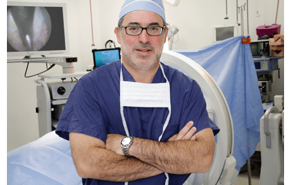
Dr. Rappard’s Los Angeles Minimally Invasive Spine Institute performs state of the art spine surgery designed to make your back pain, neck pain or sciatica go away faster. We only perform back surgery and neck surgery that is ultra-minimally invasive. We use sophisticated high resolution cameras placed through natural openings in the spine, miniaturized surgical instruments and a spine laser, all inserted through a tiny skin incision. Our minimally invasive spine surgery is done without cutting or disrupting normal muscles, ligaments or bone. As a result, your recovery is much quicker. After your surgery your body can concentrate on healing from your condition, not healing from the traumatic effects of surgery. Whether it’s for state of the art sciatica treatment, back pain or neck pain, the institute has specialized techniques that can get you better faster.
Contact us for more information about cutting edge spine care.
Tactical and Sports Athlete Program
Spine injured tactical athletes (military, law enforcement and fire) and sports athletes are unique among patients. Tactical and sports athletes have less time to recover from their spine injuries and are likely to return to high-risk activities. These nuances have led the Los Angeles Minimally Invasive Spine institute to adopt a philosophy for treating these athletes that is as unique as they are. Make the most of intensive therapy: The spine injured athlete is highly motivated and is injured in a state of high physical condition. They can make the most of an intensive therapy program that relies on exercise and conditioning, while employing typical modalities. We have partnered with therapy facilities throughout Southern California that have experience and a reputation for excellence in treating athletes. Patient athletes undergoing therapy will have their care monitored by a Medical Doctor with training and specialization in rehabilitation therapy. Therapy is closely monitored and interventions are employed so that the patient athlete has the best opportunity to progress and improve.
Share information between providers and athletes: Collaboration among professionals is basic to a patient’s care. Now, the patient athlete is included in that collaboration. The Institute utilizes a unique electronic patient portal, allowing the care records to follow the athlete on a personal electronic device. The portal not only allows the athlete to have instant access to all records, it allows every medical professional seen by the athlete to access the athlete’s records, with the athlete’s permission. The introduction of the patient portal means that the athlete knows what care providers are thinking and doing, allowing the athlete to participate in treatment decisions and to easily share information among medical providers.
Maximize recovery through a minimalist approach: This is the bulwark of our core treatment philosophy. Every athlete deserves the opportunity for a non-invasive recovery through a quality therapy program. If necessary, we perform spinal injections early during therapy so that patients can return to therapy and be successful. When therapy and injections are ineffective, care is escalated to include the unique ultra-minimally invasive procedures performed by the Institute. These special procedures preserve normal spinal motion, muscles and stability to minimize recovery time, maximize rehabilitation potential and minimize re-injury.
 Comprehensive care is continuity of care: The institute is unique in its comprehensive nature. Our program integrates physician expertise in rehabilitation medicine, spine imaging, spine injections and endoscopic spine surgery. We have partnered with one of the leading chiropractic universities in the nation and with leading sports therapy facilities throughout Southern California. The comprehensive nature of care is important; it means that an athlete can be cared for almost entirely within our system of physicians and partners, ensuring continuity of our unique athlete treatment philosophy.
The tactical and sports athlete is not the average patient. They must be recovered quickly and returned to high-risk activity. These athletes will benefit from intensive therapy, specialized physicians, patient and provider collaboration and minimalistic procedures that preserve an athlete’s strength and abilities. This comprehensive care program is now available at the Los Angeles Minimally Invasive Spine Institute for tactical and sports athletes at all levels.